Helicobacter pylori (H. pylori) is a type of bacteria. These germs can enter your body and live in your digestive tract. After many years, they can cause sores, called ulcers, in the lining of your stomach or the upper part of your small intestine. For some people, an infection can lead to stomach cancer.

Infection with H. pylori is common. About two-thirds of the world’s population has it in their bodies. For most people, it doesn’t cause ulcers or any other symptoms. If you do have problems, there are medicines that can kill the germs and help sores heal.
As more of the world gets access to clean water and sanitation, fewer people than before are getting the bacteria. With good health habits, you can protect yourself and your children from H. pylori.
How H. pylori Makes You Sick
For decades, doctors thought people got ulcers from stress, spicy foods,smoking, or other lifestyle habits. But when scientists discovered H. pylori in 1982, they found that the germs were the cause of most stomach ulcers.
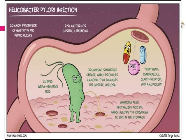
After H. pylori enters your body, it attacks the lining of your stomach, which usually protects you from the acid your body uses to digest food. Once the bacteria have done enough damage, acid can get through the lining, which leads to ulcers. These may bleed, cause infections, or keep food from moving through your digestive tract.
You can get H. pylori from food, water, or utensils. It’s more common in countries or communities that lack clean water or good sewage systems. You can also pick up the bacteria through contact with the saliva or other body fluids of infected people.
Many people get H. pylori during childhood, but adults can get it, too. The germs live in the body for years before symptoms start, but most people who have it will never get ulcers. Doctors aren’t sure why only some people get ulcers after an infection.
Symptoms
If you have an ulcer, you may feel a dull or burning pain in your belly. It may come and go, but you’ll probably feel it most when your stomach is empty, such as between meals or in the middle of the night. It can last for a few minutes or for hours. You may feel better after you eat, drink milk, or take an antacid.
Other signs of an ulcer include:
• Bloating
• Burping
• Not feeling hungry
• Nausea
• Vomiting
• Weight loss for no clear reason
Ulcers can bleed into your stomach or intestines, which can be dangerous to your health. Get medical help right away if you have any of these symptoms:
• Stool that is bloody, dark red, or black
• Trouble breathing
• Dizziness or fainting
• Feeling very tired for no reason
• Pale skin color
• Vomit that has blood or looks like coffee grounds
• Severe, sharp stomach pain
It’s not common, but H. pylori infection can cause stomach cancer. The disease has few symptoms at first, such as heartburn. Over time, you may notice:
• Belly pain or swelling
• Nausea
• Not feeling hungry
• Feeling full after you eat just a small amount
• Vomiting
• Weight loss for no reason
Getting a Diagnosis
If you don’t have symptoms of an ulcer, your doctor probably won’t test you for H. pylori. But if you have them now or have in the past, it’s best to get tested. Medicines like nonsteroidal anti-inflammatory drugs (NSAIDs) can also damage your stomach lining, so it’s important to find out what’s causing your symptoms so you can get the right treatment.
To start, your doctor will ask you about your medical history, your symptoms, and any medicines you take. Then she’ll give you a physical exam, including pressing on your belly to check for swelling, tenderness, or pain. You may also have undergo some invasive procedure such as colonoscopy so they can get a sample of your stomach lining and check it for presence of H. Pylori.
However, there is an easier way to get yourself tested by using readily available home test kit such as Fujibio H.Pylori Test Kit.

The Fujibio H.Pylori Test Kit detects helicobacter pylori through visual interpretation of color development on the internal strip. Anti-H. pylori antibodies are immobilized on the test region of the membrane. During testing, the specimen reacts with anti-H pylori antibodies conjugated to colored particles and precoated onto the small pad of the test. The mixture then migrates through the membrane by capillary action and interacts with regeants on the membrane. If there is sufficient H.pylori antigens in the specimen, a colored band will from at the test region of the membrane. The presence of this colored band indicates a positive result, while its absence indicates a negative result. The appearance of a colored band at the control region serve as a procedural control, indicating that the proper volume of specimen has been added and membrane wicking has occurred.
PROCEDURE
Bring tests, specimens, buffer and/or controls to room temperature (15-300 c) before use.
1.Specimen collection and pre-treatment:
1. Use clean,dry containers for specimen collection . best results will be obtained if the assay is performed within 6 hours after collection. Do not leave specimens at room temperature for prolonged periods. Specimens may be stored at 2-8o C for up to 72 hours. If specimens are to be shipped, pack them in compliance with all applicable regulations for transportation of etiological agents.
2.Unscrew and remove the dilution tube applicator. Be careful not to spill or spatter solution from te tube. Collect specimens by inserting the applicator stick into atleat 3 different site of the feces to collect approximtately 50mg of feces (equivalent to ¼ of a pea.)
3.Replace the applicator back into the tube and screw the cap tightly. Be careful not to break the tip of the dilution tube.
4.Shake the collection tube vigorously to mix the specimen and the extraction buffer.
2. Testing
1) Remove the test from its sealed pouch, and place it on a clean, level surface. Label the test with patient or control the identification. To obtain a best results, the assay should be performed within one hour.
2. Using a piece of tissue paper, break the tip of the dilution tube. Hold the tube verticallyand dispense 3 drops of solutioninto the specimen well (S) of the test device.
Avoid trapping air bubbles in the specimen well (S), and do not add any solution to the result area. As the test begins to work, color will migrate across the membrane.
3. Wait for the colored band(s) to appear. The result should be read within 15 minutes. Do not interpret the results after 20 minutes.
Note: If the specimen does not migrate due to the precense of particles, centrifuge the extracted specimens contain in the extraction buffer vial. Collect 100 ul of supernatant, dispence to the specimen well (S) of a new test device and start again, following the instructions described above.
INTERPRETATION OF THE RESULTS
Positive: Presence of two colored bands, one in the control region (C) and another in the test region (T), indicates presence of antibodies to H. pylori in specimen.
Negative: Presence of a colored band in in the control region (C) with no band in the test region (T) indicates absence of antibodies of H. pylori in the specimen
Invalid: If after 10 minutes no band is visible, or if a band appears in the test region (T) only, the result is invalid. If invalid, the assay should be repeated using a new test kit.